13 December 2020
Brain scans offer us a rare sight: A look into the mind and its mysteries. They promise answers to many questions. How do we feel pain? How do we know a face? How do we move our body?
But can brain scans really answer these questions? Many scientists are now rethinking the value of brain scan research and whether its findings are true.
Brain scan studies have been criticized for several things. Criticisms include using too few subjects and incorrectly reading results.
Researchers have also come to understand that a person's brain scan results can be different from day to day, even when all the conditions stay the same. Now they admit that brain scan findings are limited. Some are studying these limitations. Others are using different methods to study the brain.
Earlier this year, Annchen Knodt, a researcher at Duke University in North Carolina, and her team published the latest paper on this topic. It questioned the usefulness of common brain scan projects. The team looked at 60 studies of the past 10 years -- including Knodt's own studies.
Watching brains ‘light up'
The research being re-examined uses a technique called functional magnetic resonance imaging, or fMRI. Using large magnets, fMRI scans find where oxygenated blood goes when someone does an activity, such as learning new words. This gives scientists a way to measure brain activity.
Researchers first began using this technology in the early 1990s. At the time, it seemed to open a window into the human brain.
Earlier imaging methods involved examining brain activity through small devices -- called electrodes -- placed on the skull.
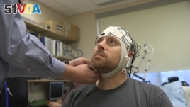
In this Nov. 20, 2019 image from video, Zach Ault is fitted with an EEG cap which uses electrodes to track the electrical activity of his brain, at the National Institutes of Health's hospital in Bethesda, Md. (AP Photo/Federica Narancio)
Another technique involved injecting radioactive material into the blood. By comparison, fMRI seemed faster and easier And the images were of higher quality.
Scientists started writing many papers about fMRI's ability to show brain activity. Reporters wrote many stories about these studies. The public wanted more.
But as years passed, troubling signs about some of the findings began to appear.
"It is a very powerful thing to show a picture of the brain," said Damian Stanley, a brain scientist at Adelphi University in New York State. However, such a powerful tool, he said, can lead to abuse and bad science. Stanley told the Associated Press that some scientists made their research seem more important than it actually was.
Another Duke brain scientist, Anita Disney, told the AP, "In the end, we probably jumped on the fMRI bandwagon a little too fast." In other words, the scientific community began supporting the technique very quickly, perhaps too quickly. But now, she said, the problems with fMRI are a concern to many scientists.
With doubts growing, many laboratories have become more careful about which imaging methods are used in brain research. And when studying the brain, there is a lot to look at. The average brain has about 177,000 kilometers of nerve fibers.
Yale University researcher Joy Hirsch wants to understand "the social brain." She studies what happens in the brain when people talk, touch, or make eye contact with each other. So, she does not use fMRI. It can only be used on one person at a time and that person must stay perfectly still inside a large scanner.
Instead, Hirsch uses another method. Laser lights hit a fiber optic cable attached to a person's head and then finds blood flow. This method lets her subjects move freely during scanning. It also permits her to study real-life socializing among several people.
Duke's Anita Disney also does not use fMRI very often. She explained that it is not accurate enough to study brain chemistry.
But not everyone is walking away from fMRI.
Some doctors depend on the method to map a patient's brain before an operation. And fMRI has proven useful for studying diseases such as schizophrenia and Alzheimer's.
Latest method for studying the brain
Now, there is a new imaging method. It is called optogenetics. This method uses light to activate neurons. It may be brain science's latest method of studying the brain. But some scientists say it is too early to know whether they will use it as a tool.
Disney noted that when a new method comes out "it is actually really difficult to get people to do the basic work of understanding its limitations."
I'm Anna Matteo.
Writers for the Associated Press reported this story. Anna Matteo adapted the story for VOA Learning English. Ashley Thompson was the editor.
________________________________________________________________
Words in This Story
scan – n. the act or process of using a special machine to see the inside of something (such as a part of the body) scan – v. to examine especially systematically with a sensing device (as a photometer or a beam of radiation)
subject – n. a person or thing that is being dealt with in a particular way
jump on a bandwagon - idiom to join an activity that has become very popular or to change your opinion to one that has become very popular so that you can share in its success
neuroscientist – n. one who studies the scientific study of nerves and especially of how nerves affect learning and behavior
nerve fiber – n. any of the processes (such as axons or dendrites) of a neuron
fiber optic cable – n. technical : the use of thin threads of glass or plastic to carry very large amounts of information in the form of light signals
activate – v. to cause (a device) to start working
basic – adj. the simplest and most important parts of something (such as a subject of study)