29 December, 2014
Researchers are now using three-dimensional, or 3D printing to create models of the human heart to help heart specialists. The heart doctors can use the models to better help patients before an operation.
Surgeons regularly use digital images to explore the heart in close detail. But no two human hearts are alike. This led Matthew Bramlet to create exact heart models from those images. Dr. Bramlet is a pediatric or children's heart expert at the University of Illinois College of Medicine. He says the 3-D models show information he cannot get any other way.
"Even when I will take the MRI and render the images, even a print heart that is sort of spinning on the screen, it's still a 2-D screen. And so what we've done with the printed models, we've pulled it out of the screen so that you can actually hold it in your hand and evaluate the anatomy for the first time in a manner that makes sense and is logical."
A 3-D printer uses images from a digital display to create a physical model of a human heart. Matthew Bramlet says doctors can use the model, in his words, "to understand the anatomy for the first time."
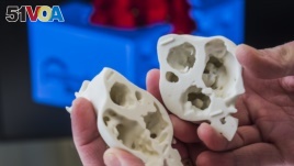
3D-printed model adds new dimension to heart surgery, allowing surgeons to see defects that might not be readily apparent in digital images. (James Carlson, Saint Francis Medical Center)
Pictures from medical tests like CAT scan or MRI are sent to a 3-D printer to create a heart in a plaster or clay form. The printer then constructs the heart, thin layer by thin layer. Dr. Bramlet says the model matches the real heart in every detail. "When we're done with the model and made our decision, we want to be able to go back to the source image and confirm those findings," he says.
Dr. Bramlet has built model hearts for different kinds of heart operations. All of the operations were successful. In his first case, digital images showed only one tiny hole in a baby's heart. But, the 3-D printed model showed several defects or problems that the baby was born with. Dr. Bramlet says those defects could not be seen easily in the images. The heart surgeon was able to change the type of surgery for the patient based on the 3-D model. He added that 3-D heart models saves time during heart operations.
"In the future, relying on that information would allow us to not even have to stop the heart to sort of go down the alternative pathway."
Kathy Magliato is a cardiac surgeon at Saint John's Health Center in Los Angeles. She welcomes the new technology. She says it could help her make better decisions before she operates on the hearts of her patients.
"The fact that I can then take this very complicated structure, which has endless possibilities of what it could look like anatomically when I open the body, and you give it to me before the operation and I can hold it in my hand and plan an operation around what I'm seeing, touching and feeling. That to me is what can potentially change the game in an operation and save lives."
Matthew Bramlet continues to research the technology. He is working with the National Institutes of Health to build a 3-D library that includes heart models and images that others can use.
I'm Jonathan Evans.
Rosanne Skirble wrote this story from Washington. Jonathan Evans wrote it for Learning English. Mario Ritter edited it.
________________________________________________________________
Words in this Story
cardiac – adj. of or relating to the heart
CAT scan – n. a picture of the inside of a part of your body that is made by a computerized machine
defect – n. a problem or fault that makes someone or something not perfect
MRI – n. magnetic resonance imaging; a method used to produce images of the inside of a person's body by means of a strong magnetic field
Now it's your turn to use these Words in this Story. In the comments section, write a sentence using one of these words and we will provide feedback on your use of vocabulary and grammar.